In the human body, the Heart is a muscular organ that is a part of the blood circulatory system. It pumps blood through blood vessels to every part of the body. We have listed down complete Human Heart Facts for Kids that will help you in learning all about the Human Heart. You are going to learn about its definition, structure, shape, location, size, weight, muscles, parts, anatomy, function, work, importance, diseases, and many other interesting facts about the Human Heart.
Human Heart Facts For Kids
What Is Human Heart
- The heart is a fundamental organ of the human body.
- It is responsible for maintaining the circulatory system of the body.
- The heart is a muscular organ that constantly pumps blood throughout the body.
- The walls of the heart are made up of strong muscles. These muscles squeeze and relax to pump blood around the body.
- The heart beats 90 times a minute in children and 70 times per minute in adults.
- The blood supplies oxygen and nutrients to different organs of the body, including the brain.
- The heart works just like two pumps and each side of the heart performs different functions.
- It is made of four chambers, called the right atrium, left atrium, right ventricle, and left ventricle.
- A thick wall of muscles, called a septum, separates the left side of the heart from the right side.
- The right side of the heart pumps blood to the lungs, while the left side of the heart pumps blood to the body.
- The functioning of the heart relies on four valves namely, mitral valve, tricuspid valve, aortic valve, and pulmonary valve.
Why Is The Human Heart Called A Double Pump
- The human heart is called a double pump because the two parts of the heart perform different pumping functions.
- The right side of the heart receives carbon dioxide-rich blood from the body and pumps it towards the lungs.
- The left side of the heart receives oxygen-rich blood from the lungs and pumps it across the rest of the body.
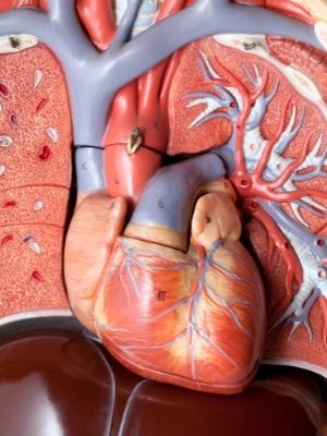
What Does A Human Heart Look Like
- The human heart is an incredible organ and it looks just like a pear or an inverted cone.
- In healthy individuals, it looks bright red, like meat. Whereas, in people with obesity, the heart looks yellow because of the yellow fatty layer.
- The heart is covered by a sac-like structure, called the pericardium. The pericardium is a double-layered membrane, which sustains the heart’s structure.
What Is The Shape Of Human Heart
- The human heart is shaped like an upside-down pear or an inverted cone.
- The base of the heart is positioned upwards and tapers down to the apex.
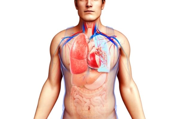
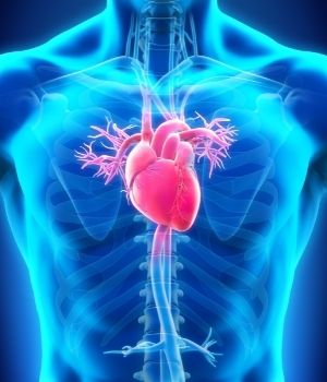
Where Is The Heart Located In The Human Body- Human Heart Location
- In the human body, the heart is located between the lungs, slightly to the left of the breastbone, and is protected by the rib cage.
- The majority of the heart’s mass is present to the left side of the body’s midline (about two-thirds).
What Is The Size Of Human Heart – Heart Size
- The human heart is the size of an adult’s fist.
- It is about 12 cm in length, 8 cm in width, and 6 cm in thickness.
- Males have slightly bigger hearts than females.
What Is The Weight Of Human Heart – Human Heart Weight
- The heart of a full-grown adult human weighs about 15 ounces (450 grams).
- The male heart has an average weight of 345 grams, while the average weight of the female heart is about 285 grams.
What Is The Heart Made Of
The heart is a muscular organ and is made of three layers of tissue:
Pericardium
- It is the outermost surface of the heart. This sac-like structure protects the heart from any external shocks, provides lubrication, and maintains its shape.
Myocardium
- It is the thick, middle layer of the heart that helps in the contraction and relaxation of the heart thus allowing blood flow.
Endocardium
- It is a thin inner layer that lines the heart chambers and also forms the surface of heart valves.
Cardiac Muscle Facts
- The cardiac muscle, also known as the heart muscle is a strained muscle present in the walls of the heart.
- It makes the middle layer of the heart, called the myocardium.
- Cardiac muscles are involuntary and work to perform different coordinated contractions to pump blood and maintain the human circulatory system.
- The contraction of the cardiac muscle forces blood out of the heart and moves it towards the lungs and body.
- It forms both the atria and the ventricles of the heart.
- Coronary arteries help cardiac muscles to deliver oxygen and blood to the body directly and also to remove any waste.
- Cardiac muscles do not diffuse like other body tissues.
- Ischemic heart disease is the most common condition that affects the cardiac muscles.
- Damage to the cardiac muscles can lead to arrhythmia and heart failure.
Parts Of The Human Heart
The human heart consists of the following parts:
Heart chambers
- The heart incorporates four chambers; two atria and two ventricles.
- The chambers work together just like a team to pump oxygen-rich blood around the body and carbon dioxide-rich blood to the lungs.
Heart valves
- The human heart has four valves; the tricuspid valve, pulmonary valve, mitral valve, and aortic valve.
- The heart valves perform the vital function of opening and closing properly to allow and control the flow of blood in the heart.
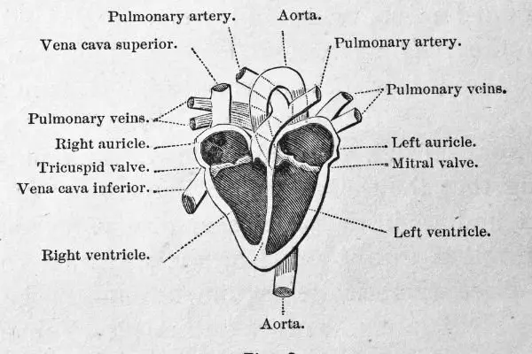
Heart vessels
- Heart vessels play an integral role in heart functions. They are; the aorta, superior vena cava, inferior vena cava, pulmonary artery pulmonary veins, and the coronary artery.
- These vessels are responsible for all the blood flow that is happening inside the body.
- The pulmonary arteries receive carbon dioxide-rich blood from the heart and send it to the lungs. The pulmonary veins transfer the oxygen-rich blood to the heart and then the aorta transports it around the rest of the body.
Heart wall
- The heart wall is composed of 3 layers; epicardium, pericardium, and endocardium.
- These layers help to protect the structure of the heart and maintain its elasticity. They also work as shock absorbers of the heart.
The conduction system
- The heart’s conduction system is a collection of specialized conduction cells and nodes.
- This system starts and coordinates the contraction of the heart muscles.
- This system works like a built-in pacemaker. It allows communication between the chambers so that they can perform coordinated functions.
Structure Of Human Heart
- The human heart has a complex structure. It pumps blood through three main divisions of the circulatory system. These systems are called; the coronary system (consisting of vessels that serve the heart), the pulmonary system (comprising the heart and lungs), and systemic (other body systems).
- The coronary circulation takes blood directly from the aorta. In the pulmonary and systemic circulation, the heart has to pump blood to the lungs or the rest of the body.
- The human heart is the size of a fist and is divided into four chambers namely, right atrium, left atrium, right ventricle, and left ventricle.
- The atria chambers are responsible for receiving the blood while the ventricular chambers are the ones that pump the blood.
- The right ventricle receives oxygen-poor blood and sends it to the lungs through the pulmonary valve.
- The left atrium, after receiving oxygen-rich blood from the lungs pumps it to the left ventricle through the mitral valve.
- The left ventricle pumps oxygen-rich blood to the rest of the body through the aortic valve.
- After that, the aortic valve closes to prevent the backflow of blood into the heart.
- The heart contracts, pushing the blood out of the heart via systole, and fills with blood again via diastole completing one cardiac cycle.
- The heart consists of cardiac cells, also known as cardiomyocytes which are responsible for the pumping of the heart.
- The heart has a built-in pacemaker that regulates and maintains the beating of the heart by using electrical signals.
Human Heart Anatomy – Gross Anatomy Of The Human Heart
- The heart performs the most physical work out of all the muscular organs in the human body.
- It is located between the ribs, slightly to the left because of its shape.
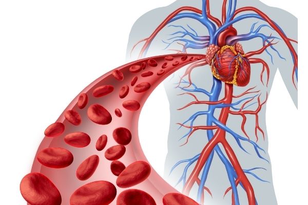
- The heart pumps blood around the body through a network of arteries and veins called the cardiovascular system. It is responsible for sending blood to the body’s cells, organs, and tissues.
- The oxygen-rich blood not only delivers oxygen to the cells but also provides important nutrients and removes carbon dioxide and other wastes.
- The system responsible for bringing blood to the heart is venules and veins. While the network that transports blood from the heart to the rest of the body consists of arteries, arterioles, and capillaries.
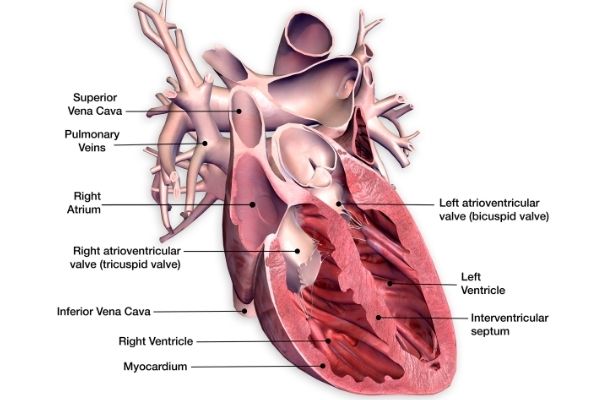
- The heart is composed of four chambers. The right atrium and left atrium are the upper two chambers while the right ventricle and left ventricle are the lower two chambers of the heart.
- It also consists of four valves: tricuspid valve, pulmonary valve, mitral valve, and aortic valve.
- All of these structures coordinate to maintain heart function.
- The superior vena cava and inferior vena cava, also known as the heart’s largest veins, bring blood to the heart, which then pumps it to the right ventricle through the tricuspid valve.
- The right ventricle pumps the blood to the lungs through the pulmonary valve, so that it can become oxygenated.
- The oxygen-rich blood is pumped to the left atrium from the lungs. The mitral valve pushes this blood to the left ventricle.
- The left ventricle transports the oxygen-rich blood to the aorta through the aortic valve which sends it across the rest of the body.
- The coronary arteries provide oxygen-rich blood to the heart.
- A complex web of nerve tissue also runs along with the heart and helps in the conduction of signals for contracting and relaxing the heart muscles.
- The heart is protected by a sac-like structure, called the pericardium. It works like a shock absorber and lubricates the heart to avoid friction.
Internal Structure Of Human Heart
- The heart’s contraction cycle sticks to a dual pattern of circulation: the pulmonary (lungs) circuit and the systemic (body) circuit.
- The two sides of the heart are divided by a structure called the septum.
- The septum that divides the atria and ventricles is called the atrioventricular septum. It consists of four openings, allowing blood flow from the atria to the ventricles and from the ventricles to the aorta.
- The heart valves are specialized structures between each of these openings that allow a one-way flow of blood. The valves between the atria and ventricles are called the tricuspid and bicuspid valves.
- The valves that lead to the aorta of the heart are called the pulmonary and the aortic valve.
- The internal cavity of the heart is separated into four chambers: right atrium, left atrium, right ventricle, and left ventricle.
- Three vessels are responsible for receiving blood in the heart: superior vena cava, inferior vena cava, and the coronary sinus.
- After acquiring blood, the high pressure moves the blood from the right atrium to the right ventricle through the tricuspid valve.
- Blood in the left atrium is acquired by four pulmonary veins and moved to the left ventricle through the bicuspid or mitral valve.
What Are The Four Chambers Of The Heart
- The heart is composed of four blood-filled areas, called chambers.
- Each side consists of two chambers, present at the top and bottom of the heart.
- The top two chambers are called atria (singular: atrium). While the bottom two chambers are called ventricles.
- Atria chambers fill with blood coming from the lungs and body. Each top side of the heart consists of an atrium, called the right atrium and left atrium.
- Ventricular chambers pump out the blood to the body and lungs. Each bottom side of the heart consists of a ventricle, called the right ventricle and left ventricle.
- The atria and ventricles work just like a team. The atria fill with blood and pass it to the ventricles which squeeze and pump that blood out of the heart.
- While the ventricles are squeezing, the atria fill with blood again for the next contraction and the cycle continues
Why Is The Left Ventricle Thicker Than The Right
- The left ventricle is thicker than the right ventricle because it pumps blood against a higher pressure as compared to the right ventricle.
- The right ventricle only pumps blood to the lungs, while the left ventricle pumps blood to the rest of the body and covers more distance.
Function Of Human Heart
The main functions of the heart include:
Pumping of blood
- The heart pumps oxygen-poor blood to the lungs, which then purifies it and sends it back to the heart. After purification, the heart pumps the oxygen-rich blood to the rest of the body.
Removal of metabolic waste
- The heart removes metabolic waste products from the blood by pumping them to the lungs.
Pumping of hormones
- Different hormones and other vital nutrients are pumped by the heart to the body for its functioning.
Maintaining blood pressure
- The heart works to maintain normal blood pressure by the process of vasodilation.
How Does The Heart Work – How The Heart Pumps Blood
- The heart works as a double pump for pumping blood.
- Each side consists of two chambers, present at the top and bottom of the heart.
- Atria are the upper chambers while ventricles are the lower chambers.
- Atria chambers fill with blood coming from the lungs and body.
- Ventricular chambers pump out the blood to the body and lungs.
- The right atrium receives oxygen-poor blood from the veins of the upper and lower body. It also receives blood from the heart itself and passes it to the right ventricle via the tricuspid valve.
- After it is filled, the blood flows from the right ventricle through the pulmonary arteries to the lungs so that it can obtain oxygen and get rid of carbon dioxide.
- The pulmonary veins pump oxygen-rich blood from the lungs to the left atrium.
- Bicuspid or mitral valves separate the chambers and blood is pumped from there to the aorta, the major artery of the body.
- The aorta transports the oxygen-rich blood to the organs and muscles of the body.
- Once blood is pumped into the aorta, the aortic valve closes to prevent the backflow of blood into the heart.
- This pattern of pumping is called double circulation and is present in all mammals.

Why Is Blood Circulation In Human Heart Called Double Circulation
- It is called double circulation because blood passes through the heart twice in one beat or one circuit.
- First, the right side of the heart pumps deoxygenated blood to the lungs where it becomes oxygenated and comes back to the heart.
- The left side then transports the freshly oxygenated blood across the body.
Human Heart Beating
- The average human heart beats 60-100 times per minute and about 100,000 times a day which means 2000 gallons of blood is pumped by the heart each day.
- The heart fills with blood before each beat, contracting the muscles to squirt the blood along.
- Contraction of the heart causes it to squeeze, causing the sinus node to send an electrical signal to the atria.
- The sinus node is the heart’s natural pacemaker, which controls the heart’s beating.
Normal Human Heart Rate – Average Human Heart Rate
- The average human heart rate for adults is between 60 to 100 beats per minute, at rest.
- According to doctors, 60 to 80 beats per minute is considered to be a normal heart rate at rest.
- A lower heart rate implies better physical and cardiovascular fitness.
- Ideally, a normal resting heart rate of 40 beats per minute is considered to be most efficient.
Human Heart Beat Per Minute
- The average human heartbeat per minute is 60 to100.
Human Heart Beat Per Day
- Every day, the human heart beats about 100,000 times.
Maximum Human Heart Rate
- The maximum human heart rate ever recorded is 480 beats per minute.
Minimum Heart Rate For Human
- The minimum heart rate for humans is about 40 to 60 beats per minute.
Major Valves Of The Human Heart
- The human heart has four major valves.
- The mitral valve and tricuspid valve, responsible for blood flow from the atria to the ventricles.
- The aortic valve and pulmonary valve, responsible for controlling blood flow out of the ventricles and to the body.
Why Are Valves Important
- Heart valves are vital for circulation and health. Each heart valve plays two important functions:
- The first function of the heart valves is to open correctly so that blood can transfer from the chamber properly.
- The second is to close properly so that the blood can move forward and not flow back to the chamber.
Human Heart Function
Aorta
- It is the largest artery of the human body, present at the top of the heart. Its function is to carry oxygen-rich blood from the left ventricle of the heart to the rest of the body.
Pulmonary artery
- The pulmonary artery is a part of pulmonary circulation that transfers oxygen and carbon dioxide to the lungs.
Pulmonary vein
- The pulmonary veins are small blood vessels that work to transfer freshly oxygenated blood from the lungs to the left atria of the heart.
Right atrium
- The main function of the right atrium is to carry deoxygenated blood to the right ventricle for purification.
Left atrium
- The left atrium holds the blood that returns from the lungs and acts as a pump to transport the blood to other parts of the heart.
Right ventricle
- The right ventricle serves to pump deoxygenated blood to the lungs.
Left ventricle
- The left ventricle is the most important chamber of the heart as it connects nearly all organ systems and pumps oxygenated blood across the body.
Human Heart Diseases
There are five major categories of heart diseases, including:
Arteriosclerosis
- Arteriosclerosis, also known as the hardening of the arteries, is a condition in which the arteries of the heart become thickened and are no longer as flexible.
- Smoking, high blood pressure, high blood glucose, and cholesterol level, all contribute to arteriosclerosis.
Atherosclerosis
- It refers to the buildup of cholesterol and fat, causing the arteries to become narrow so less blood can flow to the heart.
- Plaque buildup can burst and trigger a blood clot which may result in a stroke.
Angina
- Angina refers to a condition in which the heart is not receiving enough blood supply.
- People who have angina feel constant pain in their chest.
Heart attack
- It happens when a blood clot or some other blockage cuts the blood flow to a part of the heart.
- The blockage occurs due to a buildup of fat and cholesterol, which can rupture blood flow.
Stroke
- Stroke is experienced when part of the brain does not receive enough blood from the heart due to a clot or a burst blood vessel.
- Brain cells begin to die and cause severe brain damage, and even death.
Heart Health Facts – Healthy Heart Facts
The heart is an essential organ of our bodies. To ensure that our heart is healthy, we can look for the following signs:
Healthy heart rate
- Normal heart rate ranges from 50 to 70 beats per minute. Exercise and training can improve this number to as low as 40 beats per minute, which indicates the excellent physical condition.
Blood Pressure
- Healthy blood pressure is usually below 120/80 mm Hg. 120 measures the arterial pressure while 80 measures the pressure when the heart is relaxed.
Cholesterol level
- Maintaining a healthy level of cholesterol is vital for the body to function properly. Cholesterol levels less than 200 mg/dL are considered normal. Whereas, too much cholesterol can form blockages and can lead to stroke and heart failure.
Recovery rate
- It refers to the ability to rebound to a normal heart rate after intensive exercise. The ideal recovery rate is a drop of 20 beats per minute.
Good oral health
- Maintaining good oral health is important for preventing heart diseases. Bacteria from the mouth can enter the bloodstream and cause arterial inflammation, as well as plaque buildup.
Heart Failure Facts
- Heart failure occurs because the heart is unable to pump enough blood into the body.
- Heart failure can affect both right and left ventricles.
- The symptoms of heart failure include shortness of breath and fatigue.
- Heart failure can also be a result of other conditions such as diabetes, coronary heart disease, high blood pressure, and heart attack.
- Atrial fibrillation is the most common cause of heart failure.
- Smoking is the leading cause of heart failure as cigarette smoke contains dangerous substances that tear the lining of the arteries called the endothelium.
- Eating too much salty food also contributes to congestion of the heart.
- Alcohol consumption triggers atrial fibrillation, causing more stress to the heart.
- Ablation is a surgical procedure that can prevent heart failure.
- Exercise can also regulate and prevent heart problems as it keeps the blood pumping across the body.
- Eating healthy foods and maintaining a healthy weight is crucial to avoiding heart failure.
Fun Facts About The Heart
- The heart is the most important organ of the human body because it pumps blood to all parts of the body and keeps it working.
- It pumps oxygenated blood (which is bright red) to the body.
- The heart pumps deoxygenated blood (which is dark red) to the lungs.
- The study of the heart is called cardiology.
- Heart cancers are rare because heart cells do not divide.
- The size of the human heart is about an adult fist.
- It weighs less than 1 pound.
- The heart usually beats about 100,000 times each day.
- The heart pumps up to 2000 gallons of blood per day.
- Men have heavier hearts than women.
- The left side of the heart is slightly bigger than the right side.
- The heart takes about 60 seconds to pump blood to every cell in the body.
- The heart has four chambers and four valves.
- The heart produces a lub-dub sound when it beats, which can be heard using a stethoscope.
- The beating sound of the heart is due to the opening and closing of the valves.
- When a child’s heart beats very fast, it is called a heart murmur.
- The rhythm of the heart is controlled by an electrical system, known as the cardiac conduction system.
- The heart can continue to beat even after it is removed from the body.
- The heart of females beats faster than males.
- Heartbeat can be checked by checking the pulse. This can be done by placing two fingers on the veins of the wrist or the side of the forehead.
- People can die of a broken heart.
- Heart attacks leave scar tissue and can lead to heart failure.